Facebook
 Google
 GitHub
 Linkedin
Researchers Develop Wireless Implant Powered by Ultrasonic Waves
Researchers from Berkeley develop a tiny implant that can detect oxygen levels of tissues deep within the body.
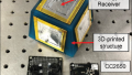
Researchers from the University of California, Berkeley (UC Berkeley), have developed an extremely small wireless implant that is powered by ultrasound and has been designed to deliver real-time measurements of tissue oxygen (O2) levels. The device would operate from the surface of the skin and take measurements from tissues deep within the body. Researchers combined the use of ultrasound technology and a sophisticated integrated circuit (IC) to create the device. The researchers originally published their work in the journal Nature Biotechnology.

The wireless, oxygen-detecting implant. Image used courtesy of UC Berkeley
Current technologies that are used to measure tissue oxygenation may use electromagnetic waves, such as infrared light, that can only penetrate the skin or organ tissues by a few centimeters. Magnetic resonance imaging can be used to provide data concerning deep tissue oxygenation, but the process of scanning can time-consuming and data cannot be provided in real-time.
Pulse oximeters can be used to measure oxygen saturation in the blood by measuring the proportion of oxygenated hemoglobin. Hemoglobin is an iron-containing component in red blood cells that can carry oxygen. The UC Berkeley team’s newly developed device can measure the amount of oxygen in tissue directly.
The New Oxygen-Detecting Implant
A professor of electrical engineering and computer sciences at UC Berkeley and a Chan Zuckerberg Biohub Investigator, Michel Maharbiz, has been designing tiny implants that operate using ultrasonic waves since 2013. The lead researcher of the current study and a postdoctoral researcher in engineering at UC Berkeley, Soner Sonmezoglu, carried on Professor Maharbiz’s work to expand the new implant’s capabilities.

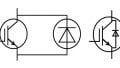
A schematic of the UC Berkeley team’s oxygen-detecting implant. Image used courtesy of UC Berkeley
The small wireless implant measures 4.5 mm long by 3 mm wide and has an oxygen sensor consisting of a micro light-emitting diode(µLED), O2-sensing film, and an optical filter. These elements are controlled by an IC. A piezo-crystal converts an electric signal from the IC into ultrasonic waves. These waves can be transmitted safely through living tissue. Piezo-crystal is a material that can generate electricity when stuck or compressed.
Potential Applications
In a news release, Sonmezoglu commented: “One potential application of this device is to monitor organ transplants, because in the months after organ transplantation, vascular complications can occur, and these complications may lead to graft dysfunction,” Sonmezoglu said. “It could be used to measure tumor hypoxia, as well, which can help doctors guide cancer radiation therapy.”
Study co-authors and pediatricians from UC, San Francisco, Jeffrey Fineman, and Emin Maltepe, believe that the device has the potential to be used for monitoring fetal development and caring for premature babies.
In the same news release, Maltepe commented: “In premature infants, for example, we frequently need to give supplemental oxygen but don’t have a reliable tissue readout of oxygen concentration.” Maltepe added: “Further miniaturized versions of this device could help us better manage oxygen exposure in our preterm infants in the intensive care nursery setting and help minimize some of the negative consequences of excessive oxygen exposure, such as retinopathy of prematurity or chronic lung disease.”
Sonmezoglu believes that the wireless implant can be improved by further miniaturization and housing the sensor. The former improvement could enable easier implantation and the latter could allow the device to remain functional in the body for longer. The optical platform in the sensor could also be modified to enable it to identify other biochemical markers in the body.
Sonmezoglu commented: “By just changing this platform that we built for the oxygen sensor, you can modify the device to measure, for example, pH, reactive oxygen species, glucose or carbon dioxide.” Sonmezoglu added: “Also, if we could modify the packaging to make it smaller, you could imagine being able to inject into the body with a needle, or through laparoscopic surgery, making the implantation even easier.”